38 heart structure with labels
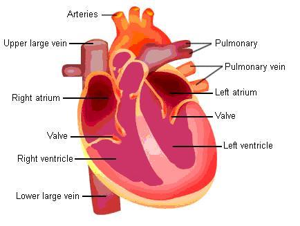
Labelling the heart — Science Learning Hub Blood transports oxygen and nutrients to the body. It is also involved in the removal of metabolic wastes. In this interactive, you can label parts of the human heart. Drag and drop the text labels onto the boxes next to the diagram. Selecting or hovering over a box will highlight each area in the diagram. Simple heart diagram | Simple heart diagram labeled - Pinterest Human heart anatomy illustrations. Hand drawn illustrations of the anatomy of the heart with labels on 4 different styles, ready to use. Line, color and texts are on different layers so they can be switched on and off as necessary. You will get: 4 EPS files 4 Ai files 4 high resolution jpeg files 4 high resolution png files L 由紀 Zapisane na szybko
Label the Heart Diagram | Quizlet Label the Heart STUDY Learn Write Test PLAY Match Created by bluesas9 Terms in this set (15) Superior Vena Cava ... Right Ventricle ... Left Atrium ... Atrioventricular/Tricuspid Valve ... Atrioventricular/Mitral Valve ... Septum ... Right Atrium ... Semi-lunar Valves ... Left Pulmonary Veins ... Right Pulmonary Veins ... Left Pulmonary Arteries

Heart structure with labels
PDF HEART - STRUCTURE - BiologyMad HEART - STRUCTURE • 4 sections Left atrium Right atrium Left ventricle Right ventricle • heart ry artery Pulmonary vein EAS the blood from he left hand side has to be pumped all around the body. • 2 lo heart Atrioventricular valves - between the atrium and the ventricles Semi-lunar valves - in the pulmonary artery and the aorta The structure of the heart - Structure and function of the heart ... Each side of the heart consists of an atrium and a ventricle which are two connected chambers. The atria (plural of atrium) are where the blood collects when it enters the heart. The ventricles... Heart Anatomy: Labeled Diagram, Structures, Function, and Blood Flow There are 4 chambers, labeled 1-4 on the diagram below. To help simplify things, we can convert the heart into a square. We will then divide that square into 4 different boxes which will represent the 4 chambers of the heart. The boxes are numbered to correlate with the labeled chambers on the cartoon diagram.
Heart structure with labels. nutritionistpro.comNutritionist Pro™ | Diet Analysis, Food Label, Menu Creation ... Designed and managed by registered dietitians for your comprehensive nutrition analysis needs. From food labels to menus to recipe calculations, Nutritionist Pro™ makes all your food science needs a simple and streamlined process. Since 1982 over 1,000,000 have relied on the Nutritionist Pro™ family of products. Structure of the Heart | The Franklin Institute The heart consists of four chambers: two atria on the top and two ventricles on the bottom. Looking at the Valentine's Day heart, the two rounded humps at the top are rounded like the top of a lower-case "a.". The bottom is shaped like a "v.". › articles › s41588/022/01090-3Genetic analysis of right heart structure and function in ... Jun 13, 2022 · Congenital heart diseases often involve maldevelopment of the evolutionarily recent right heart chamber. To gain insight into right heart structure and function, we fine-tuned deep learning models ... How to Draw the Internal Structure of the Heart (with Pictures) Coloring and Labeling 1 Color these pink: Border Left Atrium Right Atrium Pulmonary Veins 2 Color these purple: Pulmonary Artery Left Ventricle Right Ventricle 3 Color these blue: Superior Vena Cava Inferior Vena Cava 4 Color this red: Aorta 5 Make sure to label the following: Superior Vena Cava Inferior Vena Cava Pulmonary Artery Pulmonary Veins
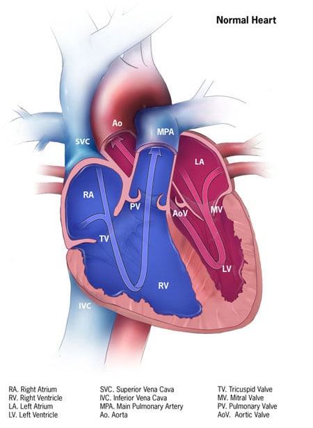
Anatomy of a Human Heart - uofmhealth The left ventricle pumps oxygen-rich blood through the aortic valve to the aorta and the rest of the body. The coronary arteries run along the surface of the heart and provide oxygen-rich blood to the heart muscle. A web of nerve tissue also runs through the heart, conducting the complex signals that govern contraction and relaxation. The Anatomy of the Heart, Its Structures, and Functions The heart wall consists of three layers: Epicardium: The outer layer of the wall of the heart. Myocardium: The muscular middle layer of the wall of the heart. Endocardium: The inner layer of the heart. Cardiac Conduction Cardiac conduction is the rate at which the heart conducts electrical impulses. Structure of the Heart | SEER Training The outer layer of the heart wall is the epicardium, the middle layer is the myocardium, and the inner layer is the endocardium. Chambers of the Heart The internal cavity of the heart is divided into four chambers: Right atrium Right ventricle Left atrium Left ventricle The two atria are thin-walled chambers that receive blood from the veins. Heart: Anatomy and Function - Cleveland Clinic Your heart walls have three layers: Endocardium: Inner layer. Myocardium: Muscular middle layer. Epicardium: Protective outer layer. The epicardium is one layer of your pericardium. The pericardium is a protective sac that covers your entire heart. It produces fluid to lubricate your heart and keep it from rubbing against other organs.
› design-templates › printHeart Diagram – 15+ Free Printable Word, Excel, EPS, PSD ... Teachers and students use the heart diagram, in biological science, to study the structure and functions of a human being’s heart. Friends and colleagues on the other hand may find this diagram template useful when it comes to sending special, personalized gifts to their family members and significant others. Download the template today, and ... Label the heart worksheets (SB6634) - SparkleBox | Human body science ... sparklebox.co.uk Label the Heart Worksheets (SB6634) A set of coloured diagrams of the heart. Includes a labelled version (simple and complex) for class discussion, as well as a worksheet for pupils to label themselves. SparkleBox 51k followers More information Label the heart worksheets (SB6634) - SparkleBox labeling the heart diagram heart diagram human label labels structure anatomy drawing interior labeled parts external clipart draw thickness internal drag drop atria varies Heart - Anterior Gross Anatomy - PurposeGames purposegames Heart Diagram - 15+ Free Printable Word, Excel, EPS, PSD Template › song-structure4 Song Structure Types to Know & When to Use Them in Your ... Apr 28, 2022 · If you were to listen to the top 10 songs on the Billboard Top 100, most or all of them would have a VCVC structure or its close cousin, Verse-Chorus-Verse-Chorus-Bridge-Chorus. So if you’re looking to become a Professional Songwriter, get comfortable writing in this structure. Examples of songs with a Verse-Chorus-Verse-Chorus structure:
Labelled Heart by abpischools - Teaching Resources - Tes
Heart Diagram for Kids - Bodytomy As you can see in the diagram of the heart, that heart is divided in four chambers, namely, right atrium, left atrium, right ventricle and left ventricle. Each of the chambers is separated by a muscle wall known as Septum. The left side of the heart receives oxygen rich blood from the lungs and pumps it out the whole body.

Heart Diagrams for Labeling and Coloring, With Reference Chart and Summary | Heart diagram ...
WebMD - Better information. Better health. The heart has four chambers: The right atrium receives blood from the veins and pumps it to the right ventricle. The right ventricle receives blood from the right atrium and pumps it to the lungs,...
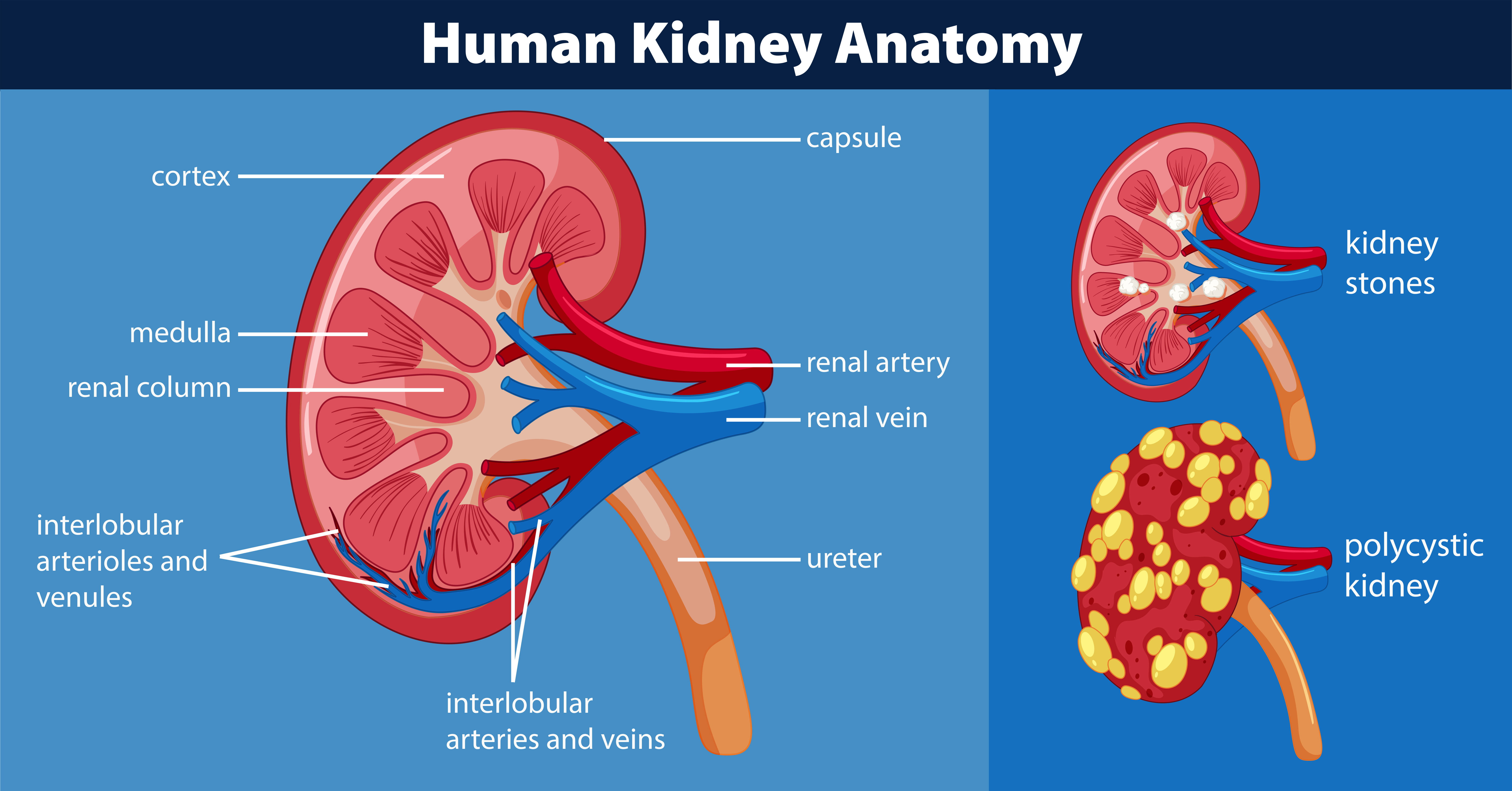
Human kidney anatomy diagram 446409 Vector Art at Vecteezy
byjus.com › biology › diagram-of-heartHeart Diagram with Labels and Detailed Explanation - BYJUS Well-Labelled Diagram of Heart The heart is made up of four chambers: The upper two chambers of the heart are called auricles. The lower two chambers of the heart are called ventricles. The heart wall is made up of three layers: The outer layer of the heart wall is called epicardium. The middle layer of the heart wall is called myocardium.
CDC - Congenital Heart Defects, Atrial Septal Defect, Graphic- NCBDDD
Structure and Function of the Heart - Medical News The heart is a muscle whose working mechanism is made possible by the many parts that operate together. The organ is divided into several chambers that take in and distribute oxygen-poor or oxygen ...
Heart Labeling (Internal)
quizlet.com › 630625176 › chapter-19-the-heart-flashChapter 19: The Heart Flashcards | Quizlet •Allows heart to beat without friction, gives it room to expand and resists excessive expansion •Parietal pericardium-tough outer, fibrous layer of connective tissue-inner serous layer •Visceral pericardium (a.k.a. epicardium of heart wall)-serous lining of sac turns inward at base of heart to cover the heart surface

Simplified Heart Labeled Decal | Shop Fathead Anatomical Images Graphics
Human Heart: Label the diagram 1 - Liveworksheets Study the figure carefully.Label the 10 parts of the human heart A-J. ID: 1781041 Language: English School subject: Biology Grade/level: 9-12 Age: 14+ Main content: Human Circulatory System Other contents: Human Heart Add to my workbooks (15) Download file pdf Embed in my website or blog

Parts Of Heart Diagram Stock Illustration - Download Image Now - iStock
Label the heart - Teaching resources - Wordwall Label the Heart Labelled diagram by Bsilver Y7 Y8 Y9 Y10 Y11 Biology First Aid Label The Heart! Labelled diagram by U32210725 Label the Heart diagram (L5) Labelled diagram by Jenniferross Y9 Biology Label the heart GCSE biology Labelled diagram by 17r2hige 10X Label the Heart diagram Labelled diagram by Kpatel1

The human egg cell explained for egg donors | Altrui
Structure of Heart (With Diagram) | Circulatory System | Human Physiology The heart is consisting of three layers: 1. Pericardium or outer covering layer: The heart lies in a double membranous sac of pericardium with serous fluid between the two layers. This is known as pericardial fluid. By its lubricating action, the heart can move freely or contracts and expands without any injury.
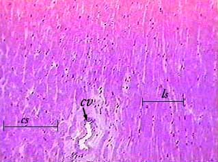
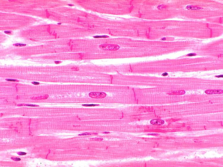
Cardiac muscle
Heart Diagram with Labels and Detailed Explanation The heart is located under the ribcage, between the lungs and above the diaphragm. It weighs about 10.5 ounces and is cone shaped in structure. It consists of the following parts: Heart Detailed Diagram Heart - Chambers There are four chambers of the heart . The upper two chambers are the auricles and the lower two are called ventricles.

Human Heart Pictures with Labels Best Of File Diagram Of the Human Heart Hug Wikimedia Mons ...
byjus.com › biology › human-heartHuman Heart - Anatomy, Functions and Facts about Heart The external structure of the heart has many blood vessels that form a network, with other major vessels emerging from within the structure. The blood vessels typically comprise the following: Veins supply deoxygenated blood to the heart via inferior and superior vena cava, and it eventually drains into the right atrium.

how to draw label diagram of heart - Science - Life Processes - 12575 | Meritnation.com
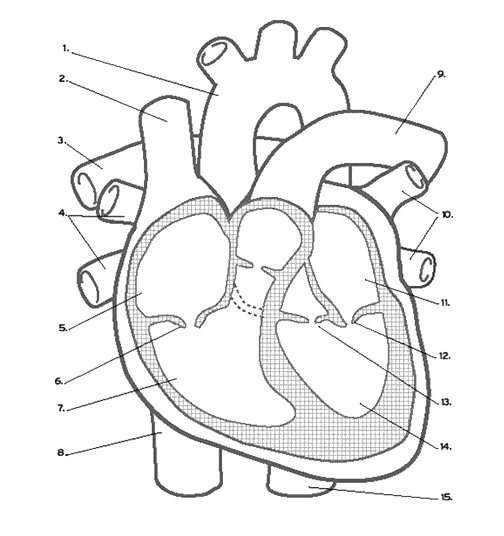
Heart Labeling Quiz: How Much You Know About Heart Labeling? Here is a Heart labeling quiz for you. The human heart is a vital organ for every human. The more healthy your heart is, the longer the chances you have of surviving, so you better take care of it. Take the following quiz to know how much you know about your heart. Questions and Answers 1. What is #1? 2. What is #2? 3. What is #3? 4. What is #4?
Tissues Flashcards | Easy Notecards
labeld diagram of the heart heart human labeled diagram label inside organ body each muscle clear chamber poster chambers cardiac Human Heart Diagram Stock Illustration - Image: 55185229 umano diagramma The Parts Or The Heart | Diabetes Inc.
![Untitled Document [www.bio.sunyorange.edu]](http://www.bio.sunyorange.edu/updated2/THINKING_EVOLUTION/physiology1/heart/c_chambers.jpg)
Untitled Document [www.bio.sunyorange.edu]
Label the heart — Science Learning Hub In this interactive, you can label parts of the human heart. Drag and drop the text labels onto the boxes next to the diagram. Selecting or hovering over a box will highlight each area in the diagram. Right ventricle Right atrium Left atrium Pulmonary artery Left ventricle Pulmonary vein Semilunar valve Vena cava Aorta Download Exercise Tweet
Know the Structures and Functions about Your Heart | New Health Advisor
The Anatomy of the Heart - Quiz 1 - Free Anatomy Quiz The circulatory system - lower body image, with blank labels attached. The circulatory system - a PDF file of the upper and lower body for printing out to use off-line. Describe and explain the function of the circulatory system - The circulatory system consists of the heart, the blood vessels (veins, arteries, and capillaries), and the blood.
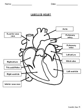
Labelled Heart by Scientific Stars | Teachers Pay Teachers
Label the HEART | Circulatory System Quiz - Quizizz True or False: Blood flows in the following sequence in the heart: Vena cava, right atrium, right ventricle, pulmonary artery, lungs, pulmonary veins, left atrium, left ventricle, aorta. Q. True or False: There are four chambers in the heart. Q. Place the pathway of blood through the heart in the correct sequence. Q.
Post a Comment for "38 heart structure with labels"