38 external structure of the heart with labels
Heart Anatomy - External - The Biology Corner Heart - External Anatomy. View of the vental surface of the heart. You can identify the front of the heart by locating the interventricular sulcus and the large pulmonary artery. The flaps on the front that cover the two atria are called the auricles. The pulmonary artery is the most anterior of the vessels and has thin walls. The blue pencil ... The Anatomy of the Heart, Its Structures, and Functions The heart is the organ that helps supply blood and oxygen to all parts of the body. It is divided by a partition (or septum) into two halves. The halves are, in turn, divided into four chambers. The heart is situated within the chest cavity and surrounded by a fluid-filled sac called the pericardium. This amazing muscle produces electrical ...
Heart Diagram with Labels and Detailed Explanation - BYJUS Diagram of Heart. The human heart is the most crucial organ of the human body. It pumps blood from the heart to different parts of the body and back to the heart. The most common heart attack symptoms or warning signs are chest pain, breathlessness, nausea, sweating etc. The diagram of heart is beneficial for Class 10 and 12 and is frequently ...
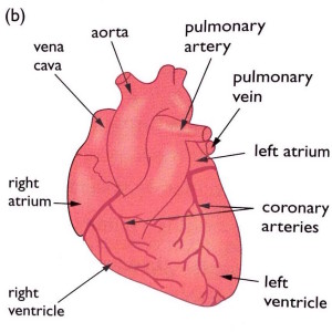
External structure of the heart with labels
The human heart (External and internal structure ... - Online Science Notes Appearance and position: Is a hollow, muscular organ (made up of involuntary cardiac muscles), roughly of the size of one's fist (12×9 cm) Average weight is about 300gm in males and 250gm in females. Is reddish-brown in color and somewhat conical in shape. Located almost in the middle of the thoracic cavity close to its front wall and ... Heart Anatomy: Labeled Diagram, Structures, Function, and Blood Flow There are 4 chambers, labeled 1-4 on the diagram below. To help simplify things, we can convert the heart into a square. We will then divide that square into 4 different boxes which will represent the 4 chambers of the heart. The boxes are numbered to correlate with the labeled chambers on the cartoon diagram. A Labeled Diagram of the Human Heart You Really Need to See The blood pumped by the heart not only provides nutrients to the body cells, but also removes the waste materials from different parts of the body. The above labeled diagram can be modified as per your requirements for kids. Heart diagram for kids can be printed out and colored, to make it easier to understand.
External structure of the heart with labels. Label the Heart - The Biology Corner Shows a picture of a heart with letters and blanks for practice with labeling the parts of the heart and tracing the flow of blood within the heart. Structure Of The Heart | A-Level Biology Revision Notes The heart is a hollow muscular organ that lies in the middle of the chest cavity. It is enclosed in the pericardium, which protects the heart and facilitates its pumping action. The heart is divided into four chambers: The two atria (auricles): these are the upper two chambers. They have thin walls which receive blood from veins. The structure of the heart - Structure and function of the heart ... The structure of the heart. If you clench your hand into a fist, this is approximately the same size as your heart. It is located in the middle of the chest and slightly towards the left. Label the heart — Science Learning Hub In this interactive, you can label parts of the human heart. Drag and drop the text labels onto the boxes next to the diagram. Selecting or hovering over a box will highlight each area in the diagram. In this interactive, you can label parts of the human heart. Drag and drop the text labels onto the boxes next to the diagram.
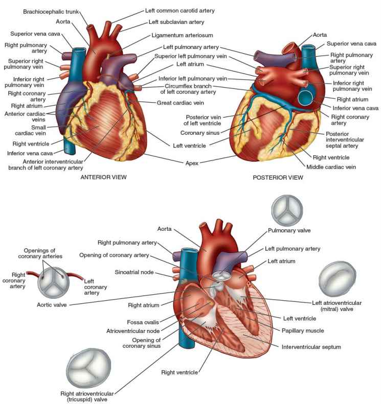
Structure of the Heart | The Franklin Institute The two largest veins that carry blood into the heart are the superior vena cava and the inferior vena cava. They are called "vena cava" because they are the "heart's veins." The superior is located near the top of the heart. The inferior is located beneath the superior. A wall called a septum, separates the right and left sides of the heart. Diagram of Human Heart and Blood Circulation in It Four Chambers of the Heart and Blood Circulation. The shape of the human heart is like an upside-down pear, weighing between 7-15 ounces, and is little larger than the size of the fist. It is located between the lungs, in the middle of the chest, behind and slightly to the left of the breast bone. The heart, one of the most significant organs ... Solved -labeling Activity: External Anatomy of the Sheep - Chegg Anatomy and Physiology. Anatomy and Physiology questions and answers. -labeling Activity: External Anatomy of the Sheep Heart Part A Drag the labels to the appropriate location in the figure. Reset Help Lolt ventric Pulmonary trunk Lolt atrium Lohtaude Right trum Posterior Interventricular sules = Pulmonary veins Art Right vorticle Anterior ... Heart Anatomy Labeling Game - PurposeGames.com This is an online quiz called Heart Anatomy Labeling Game. There is a printable worksheet available for download here so you can take the quiz with pen and paper. Your Skills & Rank. Total Points. 0. Get started! Today's Rank--0. Today 's Points. One of us! Game Points. 19. You need to get 100% to score the 19 points available.
external structure of heart How would you label the structures (both external and internal) of a we have 8 Pictures about How would you label the structures (both external and internal) of a like How would you label the structures (both external and internal) of a, Blood Vessels Flashcards | Easy Notecards and also Toad Dissection (how to/how not to) - YouTube. External anterior heart labeling Quiz - PurposeGames.com This is an online quiz called External anterior heart labeling. There is a printable worksheet available for download here so you can take the quiz with pen and paper. byjus.com › biology › human-heartIntroduction to the Human Heart - BYJUS All vertebrates, including humans, possess this type of circulation. The external structure of the heart has many blood vessels that form a network, with other major vessels emerging from within the structure. ... Label the Heart Diagram below: Practice your understanding of the heart structure. Drag and drop the correct labels to the boxes ... en.wikipedia.org › wiki › The_TenorsThe Tenors - Wikipedia The Tenors (formerly known as The Canadian Tenors) are a vocal group consisting of Victor Micallef, Fraser Walters, and Clifton Murray.They perform operatic pop music that is a mixture of classical and pop, featuring songs such as "The Prayer", Panis angelicus, and Leonard Cohen's Hallelujah.
iGCSE Biology - Gross Structure Of The Heart - BioChem Tuition
Solved Help Label the external anatomy on this posterior - Chegg Expert Answer. 100% (6 ratings) Transcribed image text: Help Label the external anatomy on this posterior view of a mammalian heart by clicking and dragging the labels to the correct location Coronary sinus Apex of heart Lert atrium Posterior interventricular branch of LCA Left pulmonary artery Left ventricle Left pulmonary veins Aortic arch.

The Heart - Biology Student
› articles › flat-vs-deep-hierarchyFlat vs. Deep Website Hierarchies - Nielsen Norman Group Nov 10, 2013 · Left: a flat site hierarchy, with few vertical levels. Right: a deep site hierarchy has the same information organized into more sublevels. Both of these site hierarchies start at the top with a single homepage, but the information below that page is organized quite differently: the website on the left has 8 major categories, but the site on the right has only 4.
Comparative Anatomy Tutorial - External Anatomy
Cardiovascular System | Anatomy of the Heart Anatomy of the Heart | Correctly Label the following External and Internal Anatomy of the Human Heart . Pericardium: The heart is placed within a fluid filled cavity called as pericardial cavity. The walls and lining of the pericardial cavity are made up of a special membrane known as the pericardium.

Pin on Classical Conversations Science
base of heart anatomy Clinical anatomy of the aortic root -- Anderson 84 (6): 670 -- Heart we have 9 Pictures about Clinical anatomy of the aortic root -- Anderson 84 (6): 670 -- Heart like PPT - Use the following terms to label external heart anatomy, Comparative Anatomy Tutorial - External Anatomy and also PPT - Use the following terms to label external heart anatomy.

The Heart | S-cool, the revision website
Heart Anatomy: size, location, coverings and layers : Anatomy & Physiology Heart Anatomy. The heart is around the size of a fist and weighs between 250-350 grams (less than a pound). Enclosed within the mediastinum, the medial cavity of the thorax, the heart extends obliquely from the second rib to the fifth intercostal space. It rests on the superior surface of the diaphragm, lies posterior to the sternum and ...

External Structure Of Heart Anatomy Diagram | MedicineBTG.com
go.drugbank.com › drugs › DB00880Chlorothiazide: Uses, Interactions, Mechanism of ... - DrugBank Chlorothiazide is indicated as adjunctive therapy in edema associated with congestive heart failure, hepatic cirrhosis, and corticosteroid and estrogen therapy. It is also indicated in the management of hypertension either as the sole therapeutic agent or to enhance the effectiveness of other antihypertensive drugs in the more severe forms of ...

The Structure & Functions of the Heart - Elite Cardiovascular Group
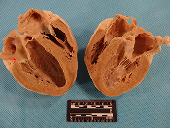
pmt.physicsandmathstutor.com › download › BiologyPractical notes - SP 2.3c Dissection of a Mammalian Heart ... The mammalian heart is a muscular pump that pushes blood around the body. It consists of four chambers and associated blood vessels . The left and right side of the heart is separated by a muscular wall, the septum . Recall the structure of the heart in the diagram below:

Aqua Fanatic: Crayfish Anatomy
Human Heart Diagram Labeled - Science Trends The pericardium is a structure that encases the heart, a sturdy structure made out of a double wall of tissue. The function of the pericardium is to hold the heart in a permanent place within the chest and give the heart protection. The outer layer of the pericardium is referred to as the parietal pericardium, and the serous pericardium is the inner layer of the structure.
Anatomy Review: The Heart
quizlet.com › 574029087 › ch-19-circulatory-systemCh. 19 Circulatory System- heart Flashcards | Quizlet 1st heart sound (S1) - The AV valves close as blood backs up against their cusps. 2nd heart sound (S2) - Blood rebounds from the closed semilunar valves and the ventricles expand. 3rd heart sound (S3) - it is thought to result from the transition from the expansion of the empty ventricles to their sudden filling with blood.

31 Blood Vessels Diagram To Label
Lesson | The Heart - External Structure | Encounter Edu Veins appear blue through the skin because of refraction but they are actually dark red. Students label the diagram of the external structure of the heart as they explore the heart and read the information (see Student Sheet found in the Lesson resources section). Students answer the questions in the scene and discuss their ideas with partners.
heart
Layers of the heart: Epicardium, myocardium, endocardium - Kenhub The myocardium is functionally the main constituent of the heart and the thickest layer of all three heart layers. It is a muscle layer that enables heart contractions. Histologically, the myocardium is comprised of cardiomyocytes.Cardiomyocytes have a single nucleus in the center of the cell, which helps to distinguish them from skeletal muscle cells that have multiple nuclei dispersed in the ...

mypicsainmarin: heart diagram with labels
quizlet.com › 630625176 › chapter-19-the-heart-flashChapter 19: The Heart Flashcards | Quizlet •Allows heart to beat without friction, gives it room to expand and resists excessive expansion •Parietal pericardium-tough outer, fibrous layer of connective tissue-inner serous layer •Visceral pericardium (a.k.a. epicardium of heart wall)-serous lining of sac turns inward at base of heart to cover the heart surface
Heart without labels
Structure of the Heart | SEER Training Structure of the Heart. The human heart is a four-chambered muscular organ, shaped and sized roughly like a man's closed fist with two-thirds of the mass to the left of midline. The heart is enclosed in a pericardial sac that is lined with the parietal layers of a serous membrane. The visceral layer of the serous membrane forms the epicardium.

Basics about Cardiovascular System : Structure and Function
Heart - External Features - Anatomy QA Location of heart: Heart lies in the middle mediastinum. 1/3rd of the heart lies to the right and 2/3rd to the left of the midline. It lies opposite to T5 - T8 vertebrae in supine position & T6 - T9 vertebrae in erect position. Dimensions of heart: Base to apex-12cm; Transversely- 8-9cm; Anteroposteriorly- 6cm.

The Cardiovascular System: Anatomy & Physiology - The Nursing Journal
Chapter 22 Heart Flashcards - Quizlet Label the structures of the pericardium in the figure. Label the external anatomy of the heart. Label the internal anatomy of the heart. Label the valves in an anterior view of the heart. Label the coronary arteries in an anterior view of the heart. Label the order that blood flows through in the heart, using the arrows as guides.
Post a Comment for "38 external structure of the heart with labels"